On the Similarity of Some Conserved Viroid Hammerhead Sequences and the Polymerase Binding Site of Methanococcus Transcription Initation complex
Sometimes, when looking for a molecule in a phylogenetic search, one looks for the conserved sequences that may have originally derived from that molecule or a progentitor. In the case of hammerhead ribozymes, one might want to use the conserved hammerhead sequences found in viroids as a starting point.
In their search for a transcription system for viroids, Pelchat, Grentier and Perrault (Biochem. (2002), 41, No. 20 p.6561) attempted to make a case, using an in vitro system, that the hammerhead sequences in PLMVD could be used as a promoter region for RNA transcription using a DNA-dependent RNA Polymerase from E. coli.
Viroids, short circles of RNA, neither code for proteins or are encapsidated by proteins. They replicate using a rolling circle mechansim that involves cleavage of multimeric long strands of RNA followed by ligation of each viroid into a circle. In a sense, replication produces the same product as transcription; RNA.
If it could be shown, as Pelchat et. al. tried, that a DNA-dependent RNA polymerase could lead to another strand of RNA than something might be said about the sequences at the site where the polymerase intiated transcription. In fact, they found that they could achieve transcription at the site of the hammerhead ribozyme on the P11 “arm” of the PLMVD viroid. As mentioned in Snippets 2, they noted that, in their in vitro system, transcription was best attained, under their conditions, if the template was in a stem loop configuration; the loop being no less than 6 nucleotides, and at the base of the loop, the A-U pair is 5’-3’, not the reverse.
Importantly, after comparing many viroids, they found that certain sequences were conserved in the region where they could generate transcription and these were derived from the viroid’s hammerhead ribozyme, as shown below.
|
From Biochemistry, Vol 41 No. 20, 2002,
Figure 7 |
|
|
|
|
|
|
|
|
|
|
|
|
|
|
|
|
|
|
||||||||||||||||
|
|
|
|
|
|
|
|
|
|
|
|
|
|
|
|
|
|
|
|
|
|
|
|
|
|
|
|
|
|
|
|
|
|
|
|
|
|
|
|
N |
A |
N |
|
|
|
|
|
|
|
|
|
|
|
|
|
|
|
|
|
|
|
|
|
|
|
|
|
|
|
|
|
|
|
|
N |
|
|
|
N |
|
|
|
|
|
|
N |
|
N |
|
|
|
|
|
|
|
|
N |
|
|
|
|
|
|
|
|
|
|
|
|
|
N |
|
|
|
|
A |
G |
U |
U |
N |
N |
|
N |
|
N |
N |
N |
N |
N |
N |
A |
N |
|
N |
N |
N |
U |
C |
N |
U |
C |
A |
G |
|
|
|
N |
|
|
|
N |
U |
C |
A |
A |
N' |
N' |
|
N' |
|
N' |
N' |
N' |
N' |
N' |
N' |
U |
N' |
|
N' |
N' |
N' |
A |
G |
N' |
A |
G |
U |
C |
|
|
|
|
N |
U |
N |
|
|
|
|
|
|
|
N |
|
N |
|
|
|
|
|
|
|
|
N |
|
|
|
|
|
|
|
|
|
|
|
|
|
|
|
|
|
|
|
|
|
|
|
|
|
|
|
|
|
|
|
|
|
|
|
|
|
|
|
|
|
|
|
|
|
|
|
|
|
|
|
|
|
Conserved nucleotides in all natural
PLMVds, ASBVd |
|
|
|
|
|
|
|
|
|
|
|
|
||||||||||||||||
|
|
|
|
|
|
|
|
|
|
|
|
|
|
|
|
|
|
|
|
|
|
|
|
|
|
|
|
|
|
|
|
|
|
|
|
|
|
|
|
A |
A |
A |
|
|
|
|
|
|
|
|
|
|
|
|
|
|
|
|
|
|
|
|
|
|
|
|
|
|
|
|
|
|
|
|
A |
|
|
|
U |
|
|
|
|
|
|
|
|
|
|
|
|
|
|
|
|
|
|
|
|
|
|
|
|
|
|
|
|
|
|
A |
|
|
|
|
|
U |
|
|
|
|
|
|
|
|
|
|
|
|
|
|
|
|
|
A |
A |
|
|
|
|
|
|
|
|
|
|
A |
|
|
|
|
|
|
A |
G |
U |
U |
C |
A |
C |
U |
|
C |
G |
|
|
U |
C |
U |
U |
|
|
U |
C |
U |
C |
3' |
|
|
|
|
|
A |
|
|
|
|
|
|
U |
C |
A |
A |
G |
U |
G |
G |
|
G |
C |
|
|
A |
G |
A |
A |
|
|
A |
G |
A |
G |
5' |
|
|
|
|
|
|
U |
|
|
|
|
U |
|
|
|
|
|
|
|
|
U |
|
|
U |
G |
|
|
|
|
G |
A |
|
|
|
|
|
|
|
|
|
|
|
|
U |
|
|
U |
|
|
|
|
|
|
|
|
|
|
|
|
|
|
|
|
|
|
|
|
|
|
|
|
|
|
|
|
|
|
|
|
|
A |
U |
|
|
|
|
|
|
|
|
|
|
|
|
|
|
|
|
|
|
|
|
|
|
|
|
|
|
|
|
|
|
|
|
|
|
|
|
ASBVd Initiation Region |
|
|
|
|
|
|
|
|
|
|
|
|
|
|
|
|
|
|
|
|
|
|||||||
|
|
|
|
|
|
|
|
|
|
|
|
|
|
|
|
|
|
|
|
|
|
|
|
|
|
|
|
|
|
|
|
|
|
|
|
|
|
|
|
|
|
|
|
|
|
|
|
|
|
|
|
|
|
|
|
|
|
|
|
|
|
|
|
|
|
|
|
|
|
|
|
There is some debate as to what the actual “transcription” site might be in viroids, and because M. Pelchat’s system is an in vitro system, it may not reflect an in vivo reality.
It might be interesting to ask if these conserved sequences are found in promoter regions in DNA. One can perhaps argue that they are, or rather they look similar to viroid conserved sequences in the following promoter/transcription binding factor regions. (The letters in bold are at similar placements to the conserved viroid ribozyme sequences and underlines are binding sites.) Below are some promoter regions, taken from the literature, which have been compared to the predicted conserved regions of the viroids shown above.
The best fit is for a sequence resembling the PLMVd transcription site is found in a paper by W. Hausner and M. Thomm entitled “Events during Initiation of Archael Transcription: Open Complex Formation and DNA-Protein Interactions. (2001) Journal of Bacteriology 183 (10) p. 3025-3031. According to their paper, transcription for the “archaeon” Methanococcus is initated by the binding of an orthologue of the TATA-box binding protein (TBF) and followed by the binding of transcription factor IIB (TFIIB). These proteins, plus RNA polymerase allow for transcription at selected promoters. The figure below is the DNAse footprint of the transcription complex and the sequence to which three proteins bind. Of note are the bases between +10 to +40 that appear to have similar conserved sequences to PLMVd in the region where transcription is initiated in M. Pelchat’s paper.
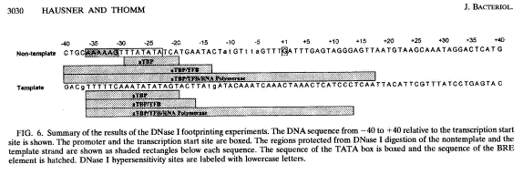
The sequence from +10 to +40 of the Methanococcus template is as
follows:
TCCCTCAATTACATTCGTTTATCCTGAGTA--C
And the sequence from PLMVd, with conserved bases highlighted, is as follows:
UNN-UCAANNNNNNNNNNNNUNNNNNGAGNAGTC
They are a very good match for the predictions for transcription initiation. As for secondary structure, it is hard to tell if a stem-loop configuration is involved or needed..
Also of interest is the sequence going 3’-5’ from +10 to –19 which looks like a hammerhead configuration, although the sequence is not totally comparable to the one from PLMVd :
TACTCAAATCAAAGTAAACATAGTATTC
What is interesting is the fact they overlap the protein binding sites of two of the proteins involved in Methanococcus trancription.
A small survey of papers saw similar sequences in the following:
Nucleotide Sequence of Promoter Region of MMTVLTR (Cell 53 371-382), (1988) The hormone regulatory unit of MMTV where an in vitro system shows the binding sites of rabbit uterine progesterone and a “receptor binding site” for glucocorticoid receptor
-180
5’ ATAAGTTTATGGTTACAAACTGttcttaaaa
TattcaaataccaatgttTgacaagaattt
________________Rabbit Uterine Progesterone Binding Site
heavier underline is binding site for glucocorticoid receptor